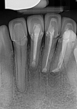
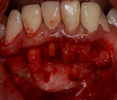
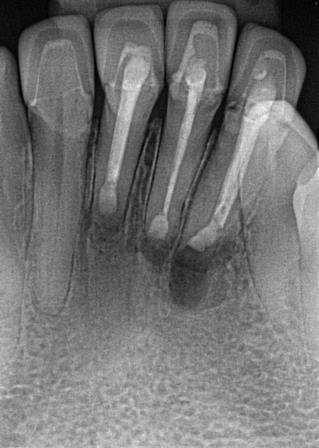
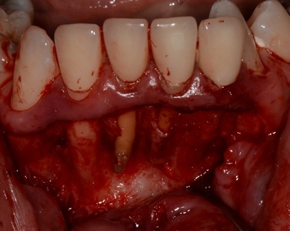
Rushing to label a tooth hopeless and proceeding to extract it can be a very costly affair for patients. Teeth with large apical pathosis should have the apical pathology managed and preferably bone regenerated prior to proceeding on to bridgework or implant based replacement. Teeth can have a regenerative potential from the PDL stem cells but also from the ability to retain soft tissue flap in a more coronal position than if the tooth was not present.
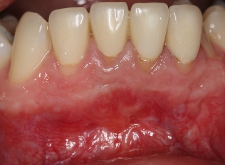
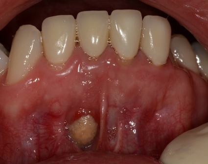
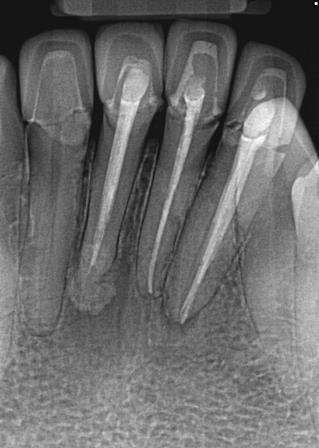
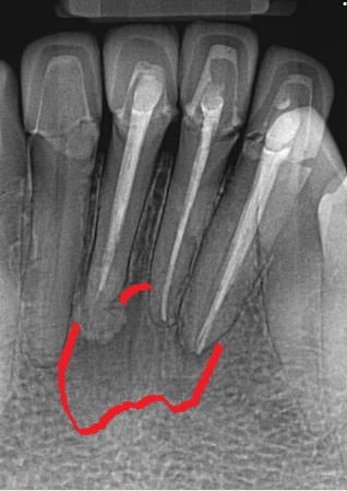
The case clearly demonstrates this. The lower incisors are buccally placed with poor endodontic obturations and poorly fitting crowns. Moreover, a large sinus resulting in dehiscence over the root of the LR1 was noted.
The options for management included:
- Accepting the situation which clearly is not appropriate given the reduction in bone volume caused by these lesions
- Considering repeating endodontic treatment and then root coverage of the LR1
- Apicectomies, with guided bone regeneration and soft tissue grafting to eliminate the dehiscence, improve the bone volume and eliminate the chronic inflammation in the site. It was made clear that the prognosis of the teeth will continue to be reduced due to the doubtful extra coronal restorations and the voids in the obturation but by increasing bone volume and improving soft tissue quality then options such as implant placement or bridge work may become more feasible.
The pictures depict the apicetomy procedure with IRM retrograde seal and Symbios (Dentsply Sirona) guide bone regeneration of the osseous defect. The soft tissues were carefully and delicately closed with 6/0 vicryl after extending the flap by considerable undermining of the lower lip. Symbios is an interesting regenerative product that is basically TCP supplied in particulate form. Dentsply encourage mixing it with blood prior to application. It was ideally suited for this case due to the excessive bleeding that occurs when the lip is undermined. Traditional paste formulation like Ethoss or Vital would be very challenging to use due to the difficulty in keeping the field dry. Dentsply recommend use of a barrier membrane with Symbyois however in this case I chose not to to avoid compromising the blood supply to the site.
The final result at 6 months was very favourable with complete healing of the dehiscence and resolution of the apical pathosis with bone regeneration. A further radiograph will be taken in 1 year to assess apical status of the incisors. Replacement with implants and further regeneration may be necessary if a relapse is noted.

